看啥推荐读物

专栏名称: 医学解剖
| 专注医学解剖,服务广大临床医生及医学生。 |
今天看啥
微信公众号rss订阅, 微信rss, 稳定的RSS源
目录
相关文章推荐

|
增删卜易 · 简体横排《全本校注初刻卜筮正宗》开始发行(多图)· 11 小时前 |


|
古典文献学微刊 · 古籍整理 | 《尚書通考》出版· 2 天前 |

|
新北方 · 五一期间出游请注意!辽宁这些景区附近将调流、限号!· 2 天前 |

|
新北方 · 小鸟轮番到电动车挡风被上薅棉花,网友:车主再 ...· 4 天前 |

推荐文章
|
|
增删卜易 · 简体横排《全本校注初刻卜筮正宗》开始发行(多图) 11 小时前 |
|
|
古典文献学微刊 · 古籍整理 | 《尚書通考》出版 2 天前 |
|
|
新北方 · 五一期间出游请注意!辽宁这些景区附近将调流、限号! 2 天前 |
|
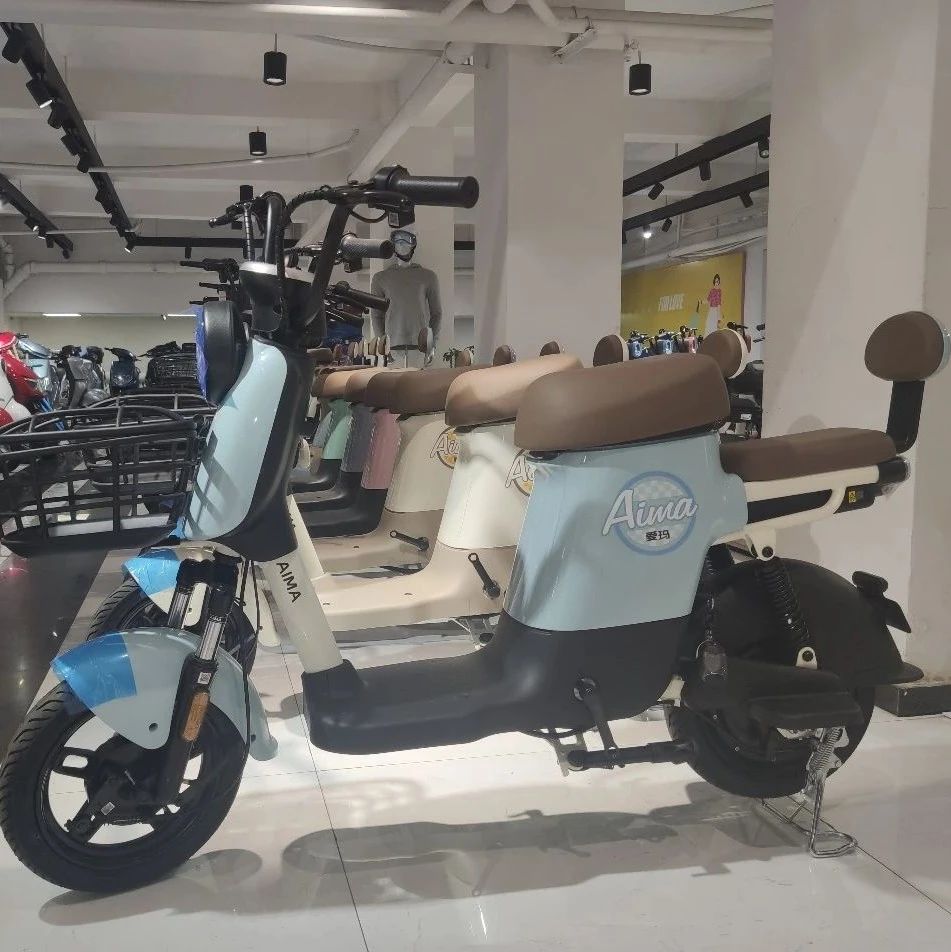
新街派 生活报 · 《黑龙江省电动车管理条例》已正式实施,如何选购合规电动车? 11 月前 |

|
深圳特区报 · 纵览深圳丨今日头版荟萃(2022.10.14) 1 年前 |
|
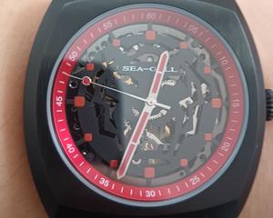
读书是一种修行 · 谈谈手表中的一些日常 2 年前 |
|
风尚云网 · 风尚云网-vue相关综合面试题(不看后悔)持续更新... 3 年前 |
|
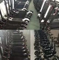
星嘉坡眼 · 转机!电动滑板车被禁,负债累累的新加坡经销商可能有救了 4 年前 |